Sommaire
- 1 How ultrasound probes actually work
- 2 The main types of ultrasound probes, and what they’re used for
- 3 Linear probes: sharp detail for structures close to the skin
- 4 Curvilinear (convex) probes: built for deeper abdominal and pelvic imaging
- 5 Phased-array (sector) probes: the small-footprint workhorse for the heart
- 6 Endocavitary probes: close-up imaging from inside the body
- 7 Micro-convex probes: a smaller option for tight spaces and small patients
- 8 “Hockey stick” probes: ultra-high frequency for tiny, superficial targets
- 9 How clinicians choose the right probe
- 10 Probe care matters more than most patients realize
- 11 Why this matters for patients, and where ultrasound is headed
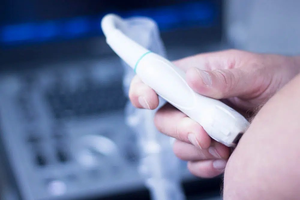
The most important piece of an ultrasound machine isn’t the screen. It’s the probe, the handheld device that sends sound waves into the body and listens for the echoes that bounce back.
Pick the wrong probe, and even a top-tier scanner can deliver fuzzy, misleading images. Pick the right one, and clinicians can spot a tiny tendon tear, track a pregnancy, or assess heart function in real time, quickly, safely, and without radiation.
Ultrasound is a workhorse of modern medicine, used everywhere from ERs and sports clinics to OB-GYN offices. But many patients, and even some non-imaging clinicians, don’t realize there are multiple probe designs, each built for a specific job and a specific depth inside the body.
How ultrasound probes actually work
Inside every probe is a line (or array) of piezoelectric crystals, materials that convert electricity into high-frequency sound waves and then convert returning echoes back into electrical signals.
Here’s the basic chain reaction: the probe fires pulses of ultrasound into tissue; those waves hit boundaries between different materials (like muscle and fluid, or soft tissue and bone) and reflect back; the probe detects the echoes; and the ultrasound system calculates how long they took to return and how strong they were. That data becomes a live image.
Key design choices, like ultrasound frequency, the number and layout of crystals, and the shape of the probe head, determine what you can see clearly and how deep you can see it.
The main types of ultrasound probes, and what they’re used for
Clinicians generally choose probes based on three things: how deep the target is, how much detail they need, and how easy it is to physically access the area. Every probe is a tradeoff between resolution (sharpness) and penetration (depth).
Linear probes: sharp detail for structures close to the skin
Linear probes have a flat face and produce a rectangular image. They typically run at higher frequencies, which means crisp detail, but limited depth.
They’re the go-to tool for superficial anatomy, including:
• Sports medicine and musculoskeletal imaging:tendons, ligaments, muscles, joints, useful for diagnosing small tears, tendinitis, and joint fluid.
• Vascular scans:carotid arteries in the neck, superficial veins in the arms and legs, clot detection, and blood-flow assessment.
• Thyroid and salivary glands:both sit close to the skin and benefit from high-resolution imaging.
• Peripheral nerves:detailed visualization for nerve blocks or suspected compression.
Upside:excellent resolution for fine structures.
Downside:limited penetration and a narrower view of deeper anatomy.
Curvilinear (convex) probes: built for deeper abdominal and pelvic imaging
Curvilinear probes have a curved face and create a wider, fan-shaped image. They usually operate at lower frequencies than linear probes, allowing sound waves to travel deeper, at the cost of some sharpness.
They’re commonly used for:
• Abdominal imaging:liver, gallbladder, pancreas, kidneys, spleen, and the abdominal aorta.
• OB-GYN:pregnancy monitoring, fetal assessment, and imaging of the uterus and ovaries.
• Urology:bladder and kidneys.
Upside:strong depth and a broad field of view.
Downside:less detailed than high-frequency linear probes for very superficial targets.
Phased-array (sector) probes: the small-footprint workhorse for the heart
Phased-array probes have a small contact surface, crucial when you’re trying to image between ribs. They generate a narrow sector image and can electronically steer the ultrasound beam, which helps capture fast-moving structures.
They’re best known for:
• Cardiology:transthoracic echocardiograms to evaluate chambers, valves, and pumping function.
• Neurology:transcranial Doppler ultrasound, which can assess blood flow in the brain through limited “windows” in the skull.
• Select abdominal or pediatric cases:when access is tight.
Upside:excellent maneuverability and strong real-time performance for motion (like a beating heart).
Downside:limited superficial field of view.
Endocavitary probes: close-up imaging from inside the body
Endocavitary probes are designed to be inserted into natural body cavities, most commonly the vagina or rectum, placing the transducer close to the target organ for high-resolution imaging.
Common uses include:
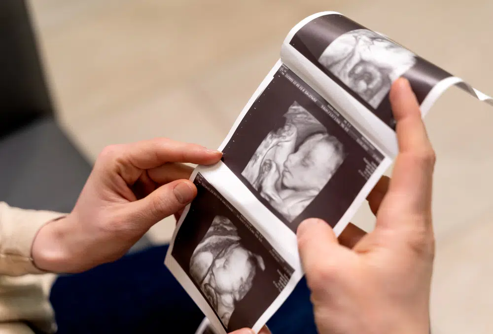
• Transvaginal ultrasound:detailed views of the uterus, ovaries, cervix, and early pregnancy.
• Transrectal ultrasound:prostate imaging and evaluation of related structures.
Upside:exceptional detail at close range.
Downside:invasive and limited field of view.
Micro-convex probes: a smaller option for tight spaces and small patients
Micro-convex probes shrink the curvilinear design into a more compact footprint, balancing depth with easier handling when space is limited.
They’re often used for:
• Pediatrics:abdominal and cardiac imaging in infants and young children.
• Veterinary medicine:especially for smaller animals.
• Hard-to-reach anatomy:when a standard curvilinear probe is too bulky.
Upside:versatile, with good penetration in a smaller package.
Downside:not as sharp as high-frequency linear probes for very superficial detail.
“Hockey stick” probes: ultra-high frequency for tiny, superficial targets
Often considered a specialized subtype of linear probe, “hockey stick” transducers are small, high-frequency tools designed for extremely superficial structures and awkward angles.
They’re used for:
• Small joints:fingers, toes, wrists.
• Superficial nerves:targeted nerve imaging for blocks or injury assessment.
• Ultra-superficial structures:skin, subcutaneous foreign bodies, and sometimes the eye.
Upside:maximum resolution and access in tight spaces.
Downside:very limited depth and a narrow viewing window.
How clinicians choose the right probe
The decision comes down to the clinical question: How deep is the structure? How much detail is needed? And what kind of access does the clinician have, between ribs, over a tender area, or inside a body cavity?
Frequency is the big lever. Higher frequency delivers sharper images but doesn’t travel as far. Lower frequency penetrates deeper but sacrifices fine detail.
Typical frequency ranges look like this:
• High-frequency linear probes:about 7–18 MHz (and “hockey stick” probes can reach roughly 10–22 MHz or higher) for superficial muscles, tendons, and nerves.
• Curvilinear probes:about 2–6 MHz for deeper abdominal and pelvic organs.
• Phased-array probes:about 2–5 MHz, optimized for cardiac motion and intercostal access.
Clinicians also think about “footprint”, the size of the probe’s contact surface. A smaller footprint can slip between ribs or fit over small joints, while a larger footprint can capture a broader view of big organs.
Probe care matters more than most patients realize
Even the best probe can underperform if it’s damaged or poorly disinfected. Drops, impacts, and harsh cleaning can degrade the probe’s materials and the crystal array inside, hurting image quality and, in some cases, raising infection-control risks.
Hospitals and clinics follow strict cleaning and disinfection protocols tailored to the probe type, especially for endocavitary probes. Proper storage, using dedicated holders instead of tossing probes onto carts, also extends lifespan and reliability.
Why this matters for patients, and where ultrasound is headed
Ultrasound probes are not interchangeable accessories. They’re specialized diagnostic tools, and choosing the right one is often the first step toward a trustworthy read, whether that’s confirming a blood clot, evaluating abdominal pain, or pinpointing a sports injury.
As probe technology keeps improving, higher resolution, better ergonomics, and more portable systems, the real win is practical: faster answers, more targeted care, and clearer images in more settings, from major hospitals to small clinics.